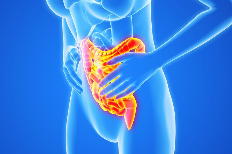
What is the procedure and how is it done?
The examination of the colon
Patients usually receive an intravenous injection of a sedative before the procedure. Sedatives help keep patients relaxed and comfortable. While patients are sedated, the doctor and medical staff monitor their vital signs.
During the colonoscopy, patients lie on their left side on the examination table. The doctor inserts a long, flexible tube with a tiny camera into the rectum and slowly guides it through the rectum and the remaining sections of the colon. The endoscope inflates the intestine with air to provide a better view for the doctor. The tiny camera transmits video images from inside the colon to a computer screen, allowing the doctor to carefully examine the tissue. The doctor may ask the patient to move at intervals for better visibility of the endoscope.
When the colonoscope reaches the end of the colon, it is slowly withdrawn, and the walls of the colon are carefully re-examined.
Biopsy and removal of colon polyps
The doctor may remove some abnormal growths from the inner surface of the intestine, known as polyps, during the colonoscopy using special tools that pass through the colonoscope. Polyps are common in adults and are usually benign at first. However, most colon cancers start as polyps, so removing them early is an effective way to prevent cancer. If bleeding occurs, the doctor can usually stop it using a special catheter, by placing clips, or with special medications that pass through the endoscope.
During the colonoscopy, the doctor may also take samples from tissues that appear abnormal. This process is called a biopsy.
 What is a colonoscopy?
 What is a colonoscopy?
